目前購物車內沒有商品
Common Name : Lajalu / Chui - Mui / Sensitive Plants
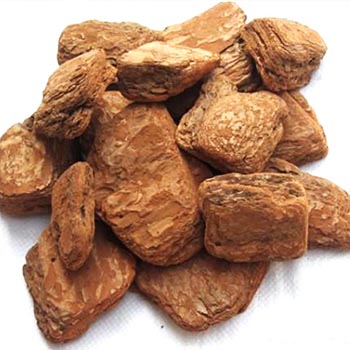
Plants Part Used : Root, Laves and Seeds

1. BOTANICAL CLASSIFICATION: Plantae Magnoliophyta Magnoliopsida Rosidae Fabales Mimosaceae
KINGDOM: Plantae
DIVISION: Magnoliophyta
CLASS: Magnoliopsida
SUBCLASS: Rosidae
ORDER: Fabales
FAMILY: Mimosaceae
Description :
Roots have characteristic 6 to 8 layers of cork cells. The secondary cortex consists of thin-walled parenchyma filled with starch granules. The cells of the cortex contain both tannin and calcium oxalate crystals.These tannin-containing cells, starch granules, crystals of calcium-oxalate, cork cells and reticulate cells are the constituents of the root powder.
Mimosa has naturally a rather high pH value, low salts and acidity and comparatively low viscosity especially in warm liquors. Like Quebracho it can be used alone or in blends, and for the reason just given it is particularly useful for blends, being capable of adjustment with other materials for many purposes. A typical blend commonly used in the U.K. Is Mimosa and a pyrogallic tannin such as Chestnut or Myrabolam. Myrabolam nuts contain pyrogallic tannins and a high proportion of organic acidity, which makes it particularly valuable for blending with a cathecol tannin like Mimosa, which is low in acidic properties. Higher natural acidity enables the degree of fixed tannin to be increased in the finished leather by decreasing the water soluble content. Pyrogallic tannin does not appreciably oxidise and improves therefore the colour fastness of Mimosa. Its light yellow colour combines well with the light pinkish colour of Mimosa.

Characteristics and Constituents :
Several studies have shown several biochemical substances involved in the contractility of the leaves. An alkaloid, mimosine has been isolated from the plant. The root extract contains 10% tannin, ash, calcium oxalate crystals and mimosine. An adrenaline like substance has been identified in the leaf extract.
On the other hand, indole-3-acetic acid (IAA) has been reported to be effective for Mimosa pudica as a leaf-opening factor. Three leaf-opening substance named potassium lespedezate, cis-p-coumaroylagmatine, and calcium 4-O-b-D-glucopyranoyl-cis-p-coumarate have been isolated from Lespedeia cuneate G. Don , Albizzia julibrissin D. and Cassia mimosoides, respectively. Based on our experiments using the leaf-opening and -closing substances, the circadian rhythm of these plants must be controlled by their internal clock which regulates a balance of concentration between the leaf-opening and closing substances.
Abstract:
Volume and conformational changes of the contractile tannin vacuoles of the abaxial motor cells of the primary pulvinus of Mimosa pudica L. parallel the seismonastic leaf movement. Since such changes in cells and organelles of animal systems are often regulated by calcium, we studied Ca2+ movement in the motor cells and tissue. By fixation with Lillie's neutral buffered formalin, followed by staining with alizarin red sulfate (ARS), calcium was localized in the tannin vacuoles of the motor cells of the primary pulvinus. After treatment with ethylenediaminetetraacetate, 8-hydroxyquinoline, and several other calcium-complexing or extracting agents, the color reaction due to alizarin red sulfonate was no longer present. By using an analytical method, it was shown that the effluent from stimulated pulvini has significantly more Ca2+ than that from unstimulated controls. Ten millimolar LaCl3 inhibits recovery of the tannin vacuole in vivo in 10 mm CaCl2 or in distilled water. Quantitative data obtained by microspectrophotometry demonstrated calcium migration during the bending movement of the primary pulvinus. In the adaxial motor cells a small amount of calcium migrates from the tannin vacuole, and calcium on the cell wall moves to the central vacuole. In the abaxial half, a large amount of calcium from the tannin vacuole moves to the central vacuole of the motor cell. It is probable that the calcium binds to the microfibrillar contents of the central vacuole. These observations support the contention that Ca2+ migrates between the surface of the tannin vacuole and the inside of the central vacuole. The recovery and maintenance of the tannin vacuole in the spherical form may play a role in maintaining turgor in the motor cells of the abaxial half of the primary pulvinus of Mimosa.
In experimental animals a crude extract from the plant showed a mild to moderate diuretic response. The total plant extract was depressant on isolated rabbit duodenum. The percent decrease in either amplitude or frequency of duodenal contractions was found to be only marginally different from that found after a similar dose of atropine sulphone. In a study of the effect of Lajjalu on regeneration of nerve in experimental animals it was seen that the plant enhances regeneration by 30-40%. The medicinal use of the plant Mimosa pudica dates back to Charaka and Sushruta. The sensitive plant is commonly used for bleeding disorders like menorrhagia, dysentery with blood and mucus, and piles. The root powder or decoction is used. The juice of freshly crushed leaves is used internally and externally in piles. A preliminary clinical trial, in 9 women with menorrhagia, exhibited promising results with relief in severity of bleeding. It is also applied externally to fissures, skin wounds and ulcers. Its action on small blood vessels is implicated in its hemostatic property.
A clinical study showed that the plant was well tolerated. It was given in the form of micronized root powder made from the dry extract. The capsules contained 500 mg and the dose was 1,000- 1,500 mg thrice a day. The acute toxicity studies in mice showed LD.
The Gelsolin/Fragmin Family Protein Identified in the Higher Plant Mimosa pudica:
Received April 24, 2001; accepted May 15, 2001
Mimosa pudica L. rapidly closes its leaves and bends its petioles downward when mechanically stimulated. It has been suggested that the actin cytoskeleton is involved in the bending motion since both cytochalasin B and phalloidin inhibit the motion. In order to clarify the mechanism by which the actin cytoskeleton functions in the motion, we attempted to find actin-modulating proteins in the M. pudica plant by DNase I-affinity column chromatography. The EGTA-eluate from the DNase I column contained proteins with apparent molecular masses of 90- and 42-kDa. The 42-kDa band consisted of two closely migrating components: the slower migrating component was actin while the faster migrating components was a distinct protein. The eluate showed an activity to sever actin filaments and to enhance the rate of polymerization of actin, both in a Ca2+-dependent manner. Microsequencing of the faster migrating 42-kDa protein revealed its similarity to proteins in the gelsolin/fragmin family. Our results provide the first biochemical evidence for the presence in a higher plant of a gelsolin/fragmin family actin-modulating protein that severs actin filament in a Ca2+-dependent manner.
Mimosa pudica L. rapidly closes its leaves and bends its petioles in response to mechanical, electrical or thermal stimulation. The bending occurs at the pulvinus, which is a motor organ located at the base of the petiole or the leaf. The response to the stimuli of parenchyma cells of the pulvinus has mainly been studied in relation to membrane potential, changes in turgor, or changes in ion flux. It is thought that the sudden loss of turgor pressure in the ventral region of the pulvinus results in the rapid bending of petioles. Previous investigations have explained the pathway of the seismonastic movement as follow: the action potential, which is generated at the stimulated site, is transmitted to the parenchyma cells of the pulvinus through the xylem and the phloem. This action potential induces the transport of ions, such as K+, Cl-, and H+, through the membranes of the parenchyma cells. The migration of these ions causes a change in the osmotic potential of the cells, which then causes a rapid change in turgor of these cells. The difference in the turgor pressure between the dorsal and ventral sides of the pulvinous results in the bending movement.
Calcium ions are considered to play a role as a second messenger in various signaling processes, not only in animal cells but also in plant cells, e.g., stomatal movements and circadian movements in nyctinastic plant. These movements are turgor-mediated, and induced by light and dark. In the parenchyma cells of the pulvinus of M. pudica, Ca ions, which are contained in both the vacuole and the apoplast, are thought to migrate to the cytoplasm during the bending movement. Thus, Ca ions may also be involved in the bending motion of Mimosa leaves and petioles.
Actin is a fundamental element of the cytoskeleton of both muscle and nonmuscle cells, and plays a crucial role in cell morphology, motility, and cytokinesis. In plant cells, the actin cytoskeleton is also involved in various phenomena such as cell growth, mitosis and cytoplasmic streaming. Fleurat-Lessard et al. have reported that cytochalasin B and phalloidin, which affect actin filaments, inhibit the bending movement of M. pudica. We have also confirmed that treatment with cytochalasin D arrests the bending movement of the Mimosa plant, while colchicine and propyzamide, both of which disrupt microtubules, have no effect (unpublished data). These results suggest that actin filaments are involved in the bending movement. However, little is known about how the actin filaments are involved in this movement. Recently, we reported that the bending motion of the Mimosa plant is correlated with reduced tyrosine phosphorylation of actin in the puluvinus. Furthermore, phenylarsine oxide, a specific inhibitor of protein-tyrosine phosphatase, inhibits both the dephosphorylation of actin and the bending of the petiole. The decrease in the level of actin phosphorylation may alter the assembly properties of actin, and then regulate the organization of the actin cytoskeleton during bending.
Other factors that may regulate the actin cytoskeleton during the bending movement are actin-modulating proteins. It is possible that these proteins change the organization of the actin cytoskeleton in parenchyma cells of the pulvinus in response to intracellular signals, which leads to the bending movement. While various kinds of actin-modulating proteins have been identified in yeasts invertebrates, and vertebrates, only a few have been identified in higher plants. Thus, it would be valuable to find novel actin-modulating proteins in higher plants in order to reveal the pathway that regulates the actin cytoskeleton during the bending movement.
In this study, we searched for actin-modulating proteins in the Mimosa plant. We obtained a fraction containing an F-actin-severing activity by DNase I affinity column chromatography. The main component of this fraction has an amino acid sequence homologous to gelsolin/fragmin family proteins.
MATERIALS AND METHODS
Plant Material:
Mimosa pudica L. was grown in a greenhouse. We used approximately 2-week-old plants to obtain tissue extracts. The plants were fully grown to be able to respond to various stimuli.
Affinity Chromatography with a DNase I Column
DNase I (Boehringer Mannheim, grade II, Federal Republic of Germany) was coupled to Affi-Gel 10 (Bio-Rad Labs., Richmond, CA, USA) according to the manufacturer's instructions.
Whole M. pudica plants were washed with de-ionized water and homogenized in 0.1 M Tris-HCl (pH 8.0), 0.4 M sorbitol, 32 mg/ml polyvinyl pyrrolidone, 0.5 mM CaCl2, 50 mM NaF, 0.5 mM ATP, 5 mM 2-mercaptoethanol (2-ME), 10% (v/v) glycerol, 10 mg/ml leupeptin, 10 mg/ml pepstatin, and 0.05% (w/v) NaN3 with a glass homogenizer. The homogenate was centrifuged at 10,000 xg for 40 min. The supernatant was filtered through a 0.45 mm membrane filter and then applied to a DNase I column pre-equilibrated with 0.1 M Tris-HCl (pH 8.0) and 0.5 mM CaCl2. The column was then washed with 50 column volumes of 0.1 M Tris-HCl (pH 7.5), 0.5 mM CaCl2, 0.1 mM ATP, and 10% glycerol. Bound proteins were eluted successively with 0.1 M Tris-HCl (pH 7.5), 5 mM EGTA, 0.1 mM ATP, 10 mg/ml leupeptin, 10 mg/ml pepstatin, 0.05% NaN3, and the same solution containing 3 M urea. Temperatures were kept at 4°C throughout the above procedures. After analysis by SDS-PAGE, the EGTA-eluate containing a 90-kDa protein and 42-kDa doublet-proteins was pooled and used for the following assays.
Preparation of Actin:
Rabbit skeletal muscle G-actin was prepared by the method of Spudich and Watt (17), and further purified by gel filtration on a Sephadex G-100 column pre-equilibrated with 2 mM Tris-HCl (pH 8.0), 0.1 mM ATP, 0.1 mM CaCl2, and 1 mM 2-ME. F-actin was obtained by the addition of 0.1 M KCl to the G-actin solution, followed by incubation for 3 h at 20°C.
Sedimentation Analysis of the EGTA-Fraction with F-Actin:
The EGTA-eluate was concentrated with Centricon 10 (Amicon, Beverly, MA, USA) and diluted ten-fold with 5 mM Hepes-KOH (pH 7.2), 0.1 mM DTT, 50 mM MgCl2, 0.1 mM ATP, 10 mg/ml leupeptin, 10 mg/ml pepstatin, and 0.05% NaN3. This procedure was repeated two more times and the sample was finally concentrated to an appropriate protein concentration. The concentrated EGTA-eluate was mixed with F-actin (final concentrations: EGTA-eluate, 2 mg/ml; F-actin, 0.2 mg/ml) and incubated for 30 min at 20°C in the presence of 0.5 mM CaCl2 or 0.5 mM EGTA. Finally the mixture was centrifuged for 2 h at 130,000 xg and the resultant supernatant and pellet fractions were analyzed by SDS-PAGE. The gel was first stained with Coomassie Brilliant Blue and then digitally photographed using a CCD camera. NIH Image software (National Institutes of Health, Bethesda, MD) was used for both image capturing and quantitation of bands on the gel.
Explained the mechanism behind the sensitive leaf curl of mimosa in terms of change in turgor pressure. I though current thinking was that it involved contractile proteins in the cytoskeleton:
Movement of leaves and leaflets in the mimosa-family is controlled by a leaf-moving motor organ called the pulvinus, located at the base of the petiole of the leaf. Changes in osmotic volume and turgor of the motor cells within the pulvinus result from the movement of ions (chiefly potassium and chloride) into and out of the cells.Signals causing leaf unfolding cause cell shrinking in the top (adaxial, flexor) one-half of the pulvinus and swelling in the bottom (abaxial, extensor) one-half. Signals causing leaf folding cause the reverse responses. In both cell types, potassium ions are released passively from the shrinking cell into the apoplast. In both cell types, the "shrinking signaling" also results in the formation of 1,4,5-inositol trisphosphate, which is a second messenger in the phosphoinositide (PI) cell signalling cascade. Calcium ions may also have a role in this regulation, but current evidence is unclear beyond this.
Kameyama et al. in 2000 (Nature 407:37) suggested that the turgor changes still occur but that the molecular process of bending may be due to decreased actin tyrosine-phosphorylation in the pulvinus. Inhibitors of the cytoskeleton such as cytochalasin B and phalloidin, also prevent these movements. Tthe same process may be associated with stomatal movements.
Actin is a protein that can form filaments which (in combination with other proteins) have the ability to contract. As such, it is an important element of the cytoskeleton of both muscle and nonmuscle cells, as well as playing a crucial role in cell morphology,motility, and cytokinesis. In plant cells, the actin cytoskeleton is also involved in various phenomena such as cell growth, mitosis and cytoplasmic streaming. Studies (Fleurat-Lessard et al., 1988) using inhibitors (cytochalasin B and phalloidin) which affect actin filament formation, also prevent the bending movement of Mimosa leaves suggesting that actin filaments are involved (in addition to the change in turgor pressure). In the most recent report (Yamashiro et al. June 2001) a protein from Mimosa that severs actin filaments has been isolated (interestingly, this is calcium ion dependent) and this may be involved in actin/cytoskeleton regulation.
So, with the best evidence based on inhibitor studies (in 1988), actin involvement in leaf movement, whilst very possible, is far from proven.

